Harness the Polypill Concept for a Broader Set of Indications

Harness the Polypill Concept for a Broader Set of Indications
(or preventive use)

“Combining renin-angiotensin system blockade and sodium-glucose cotransporter-2 inhibition in experimental diabetes results in synergistic beneficial effects”

https://pubmed.ncbi.nlm.nih.gov/38088400/

I’ve seen interesting advocacy for use of a polypill in cardiovascular disease, even heart failure with reduced ejection fraction; usually 3 or 4 meds including a choice of beta-blockers, angiotensin converting enzyme inhibitors, angiotensin receptor blockers, mineralocorticoid receptor antagonists, all in low effective doses. Generally such combinations are reported well-tolerated.

E.g., “Polypill Strategy for Heart Failure With Reduced Ejection Fraction”

https://ctv.veeva.com/study/polypill-strategy-for-heart-failure-with-reduced-ejection-fraction

Omitting the beta-blocker and mineralocorticoid receptor antagonists as unneeded except in heart failure cases, consider a polypill based on a sodium-glucose cotransporter-2 inhibitor and an angiotensin receptor blocker. Both these med classes are in wide use, well tolerated, and effective in their usual indications. Underlying their canonical mechanisms, benefits for mitochondrial function appear involved, most simply through SGLT2i inducing mild ketosis and ARB leading to AMPK activation or other effects on mitochondrial dynamics (also below). Prevalant conditions that may benefit from this approach include DM II including the risk of cardiovascular and renal complications, MASH and related liver fibrosis, other cardiovascular and renal diseases.

I’d wager that optionally adding in acarbose, another generally safe and well-tolerated med, separately administered with 1-2 meals a day, would result in a gentle weight loss. The acarbose intestinal glucosidase inhibition would be independent of the SGLT2i glycosuria effect, but compatible and complementary to further lower glucose availability in circulation, prompting compensatory lipolysis and liver gluconeogenesis and so gradually decreasing the adipose burden in patients who may need this.

Modulation of Mitochondrial Dynamics by the Angiotensin System in Dopaminergic Neurons and Microglia

https://www.aginganddisease.org/EN/10.14336/AD.2024.0981

mobilemito.blog

Essentially complete transfection of recipient cell mitochondria, by donor cell mitochondria

Source:

Enhancing radiation-induced reactive oxygen species generation through mitochondrial transplantation in human glioblastoma

Scientific Reports | (2025) 15:7618

http://pubmed.ncbi.nlm.nih.gov/40038364

Excerpt from abstract:

“Glioblastoma (GBM) is the most aggressive primary brain malignancy in adults, with high recurrence rates and resistance to standard therapies. This study explores mitochondrial transplantation as a novel method to enhance the radiobiological effect (RBE) of ionizing radiation (IR) by increasing mitochondrial density in GBM cells, potentially boosting reactive oxygen species (ROS) production and promoting radiation-induced cell death. Using cell-penetrating peptides (CPPs), mitochondria were transplanted into GBM cell lines U3035 and U3046. Transplanted mitochondria were successfully incorporated into recipient cells, increasing mitochondrial density significantly. Mitochondrial chimeric cells demonstrated enhanced ROS generation post-irradiation, as evidenced by increased electron paramagnetic resonance (EPR) signal intensity and fluorescent ROS assays. The transplanted mitochondria retained functionality and viability for up to 14 days, with mitochondrial DNA (mtDNA) sequencing confirming high transfection and retention rates. Notably, mitochondrial transplantation was feasible…”

(Emphasis added.)

Interpretation, any errors my own:

  1. Human brain tumor-derived cells in culture were selected for use in a study examining a hypothesis that increasing mitochondria numbers in the cancer cells would result in greater vulnerability to cell death after exposure to radiation. If the hypothesis was supported, this could point toward a way to improve the radiation therapy tumor response in human brain cancers.
  2. The cancer-killing mechanism proposed for examination was simply that increasing the number of radiation-damaged mitochondria within cancer cells would hasten their death after the therapy. Such damaged mitochondria would function poorly and so produce greater amounts of toxic products, such as reactive oxygen species (ROS).
  3. The method chosen to increase mitochondrial numbers in the tumor cells was isolation of native mitochondria from cell lines of the same or an alternate similar type; after tagging these with stains for visibility, and with peptides known to promote entry into cells, the fresh mitochondria were added to the cancer cells in culture. Mitochondrial entry into the tumor cells was expected, as has been shown in many studies of mitochondria transfer.
  4. The outcome examined for was a significant increase in the ROS production in mitochondria-treated cells, compared with control, untreated cultures.The study outcome measures supported the hypothesis; the experimental cells that had their mitochondrial numbers increased by the transfer, and did produce significantly higher amounts of ROS. This suggests continued exploration of the transfer technique, as clinical use may be possible after further study.

Thought-provoking additional findings:

The mitochondria that were transferred into cells, either from the same line or an alternative line, showed persistence and function in their new cells. They “took root”.

Because mitochondria are mobile within and between cells, some degree of merging, called fusion, between native and newly entered mitochondria was expected. Fusion makes possible the exchange of genetic material among different individual mitochondria, aided by their resident partial genomes being bacteria-like, small, circular, lacking a protective membrane, and present in multiple copies. This fusion and genomic mixing was demonstrated, by sequencing of the mitochondrial genomes in the treated cells and finding sequences from both types of cell lines present in frequent combination with one another.

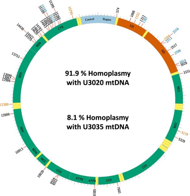
In those cells that had received mitochondria from an alternate line (“chimeric” cells, bearing two lineages of mitochondrial genomes) the sequencing made it possible to distinguish between genomic sequences that were original, and those sequences that had originated from the transferred alternate line of mitochondria. Fusions of new and original genetic elements, a virus-like process called transfection, were indeed found. In fact, the newly entered mitochondrial genome sequences were shown to nearly completely replace the originals in the chimeric cells. The group has illustrated this finding in the colorful chart below.

www.ncbi.nlm.nih.gov/core/lw/2.0/html/tileshop_pmc/tileshop_pmc_inline.html

Here in the chart, “homoplasmy” means “these DNA sequences fairly uniformly match up with reference sequences”. (And heteroplasmy is when sequences do not match up in this way.) U3020 is the mitochondrial donor cell line, U3035 is the recipient. And the long designation U3035Mt20-X3 refers to those recipient cells, treated with a higher dose of the U3020 mitochondria.

“…it was found that the DNA sequence in the 14-day post-transplant U3035Mt20-X3 chimeras was most similar to the donor U3020 line and had only retained a small proportion of the recipient mtDNA sequence.”

The donor DNA sequences had substantially replaced those of the recipient’s original genome. Perhaps general similarity of these human cancer-derived cell lines helped here.

Within this group’s model, this last finding is a demonstration of a process I have long wished to know more about. I can’t say how applicable the result is beyond this model, or how generalizable these findings are.

Why do I see potential in this finding? Think about the discussions we are beginning to hear concerning replacement of human mitochondria as a therapeutic method comtemplated for many conditions. This idea assumes that the native mitochondria in any of us exist with varying degrees of damage accumulated in the process of their constant biochemical activity. This damage does specifically include mitochondrial DNA deletions, missing or mistaken base pairs, resulting in errors in the ribosomes or proteins produced, and so leading to impairment of function. Long exposure to the active biochemistry of the mitochondrion leads to these, causing a loss of information that is necessary not only for transcribing mitochondrial proteins, but also the production of new mitochondria, or mitochondrial “biogenesis”.

This biogenesis is a cooperative readout of mitochondrial genes that have become located over time in the protected, membrane-enclosed cell nucleus, along with genes present in the smaller and more fragile remnant mitochondrial genome. Gene sequences from both these locations are necessary, and the small mitochondrial genome that remains does encode critical genes for energy production and other essential functions. Together these gene transcripts make possible assembly of new replacement mitochondria. But as DNA deletions will accumulate with time in the mitochondrial genomes in a nearly universal process, the mitochondria resulting from biogenesis will unavoidably have corresponding impairments and loss of efficiency. Credible theories of aging have been built around these observations.

The goal of restoring competent mitochondria in an ill patient, or in any of us as we age and suffer impairments, may be reached at some near-future point, given progress in techniques of harvesting mitochondria from various types of cells in culture. Mitochondria newly harvested from cell cultures should initially show a minimum of damage and deletions.

Metaphors used to make this proposed restoration process understandable have often originated from the well-known “powerhouse of the cell” image, a largely true but very incomplete description. And so these ideas are communicated in terms of reconnecting power sources or replacing batteries. There may be a certain aspect of true representation here; research findings have often shown that transferred mitochondria may arrive in recipient cells competent and able to function energetically.

But the mitochondria also store genomic information necessary for their function and continued biogenesis. When through illness or aging, mitochondrial DNA deletions have accumulated, will transferred more complete mitochondria be able to make up for these losses? Are new mitochondria imported into cells able to effectively lend their more intact genomic information into the recipient mitochondrial network through fusion, with subsequent replication of the mingled DNA favoring restoration of the information missing through deletions present in the recipient cell? If that were a possible outcome, biogenesis could again produce complete and competent mitochondria in ongoing fashion.

We can’t yet know for certain about a hoped-for therapeutic genomic restoration from this one study in cultured human cancer cells; but these findings do show a near-complete replacement of the recipient mitochondrial genome by sequences from the donor cells. A demonstration of what may be possible for therapeutic use in disorders of mitochondrial function, including many chronic diseases and pathologies of aging.

Another thought-provoking finding in the mitochondrial transfer field; there have been very many in recent years. I feel that increased interest and investment in this area will lead to advances in clinical medicine, in the near future.

You just can’t get more proximal to the biochemical action of our kind of life, than mitochondria.

CGM overestimates glucose in comparison to capillary sampling

I have heard that continuous glucose monitors (CGM) are becoming popular among individuals interested in their responses to diet, and their health risks.

In relation, this appears important:

A pre-proof from the American Journal of Clinical Nutrition

https://ajcn.nutrition.org/article/S0002-9165(25)00092-9/fulltext

Note, only one brand and model of CGM was used in the study. Significant variations in CGM values among different individuals tested under similar conditions were also observed. Some caution in generalizing findings is justified.

Thebgroup reports a mean overestimation by CGM versus capillary sampling as 0.9 mm/l, which is 16.2 mg/dl.

And this contributed to larger CGM calculated values for area, in area-under-the-curve calculations; e.g. the duration of hyperglycemia after a glucose challenge or a meal.

Conversion of mmol/ L into mg/dL is x18. Clear circles are CGM values, red are those values adjusted for difference relative to capillary samples.

“CGM-estimated fasting and postprandial glucose concentrations were(mean±SD) 0.9±0.6 and 0.9±0.5 mmol/L higher than capillary estimates, respectively(both, p<0.001).”

“The increase in glucose concentrations measured with CGM versus the criterion method resulted in a >3.8-fold increase in the time spent above 7.8 mmol/L…”

“CGM overestimated glycemic responses in numerous contexts. At times this can mischaracterize the GI [glycemic index]. In addition, there is inter-individual heterogeneity of the accuracy of CGM to estimate fasting glucose concentrations. Correction for this difference reduces, but does not eliminate, postprandial overestimate of glycemia by CGM. Caution should be applied when inferring absolute or relative glycemic responses to foods using CGM, and capillary sampling should be prioritised for accurate quantification of glycemic response.”

Mitochondria are Mobile

Saturday, February 22, 2025

Over generations or during a lifespan, whole and individual, or in fragments 

  • Early in life’s origins, as now, there were many different lineages of living cells with each bearing individual adaptations. Keeping alive requires resources to be brought into a cell; some cells evolved a capacity to engulf not only useful nutrients but also absorb other cells. A cell engulfed by another would likely be broken down for resources unless it had evolved effective defenses.
  •  At this microscopic scale, living matter may show unexpected persistence. Sometimes an engulfed cell could defend against breakdown and partially survive, continuing its life as a passenger, perhaps a parasite, or better, a valuable symbiont. Evidence for this process as it happened then and continues now, is visible under a microscope.
  • When this survival by symbiosis merged cells with different but compatible abilities, a new form of cell could result, opening a trail for new kinds of life to appear and expand.
  • Our bodies, and much of the life we see around us, are built of cells that were originally such a new lineage, one bearing a fortunate combination of abilities, with its living components evolving adaptations to each other. That cooperation is ongoing then and now, with the symbiotic partners mutually changing over time.
  • Mitochondria became a symbiotic partner in cells in this way. They were once a type of bacterial life which had evolved a highly efficient method of producing useful energy from nutrients. Cells engulfing mitochondria that could keep this new passenger’s energetic function harnessed and regulated for mutual purposes benefited tremendously. The surplus energy acquired relaxed former limits on growth and genetic information capacity in the new lineage of cells.

Cell Nucleus, Mitochondrial Nucleoid

  • The dual sets of DNA in this new lineage became mutually adapted over time. The larger genetic set, the cell nucleus, was protected by a membrane and packaged with specialized protective proteins, while most of the original mitochondrial gene set was copied over into the nucleus during this long process of adaptation. The DNA remaining behind within the mitochondria reflects its bacterial origin: it is circular, smaller, and lacks a protective membrane. There are usually several copies of this remnant genome within each mitochondrion, and many mitochondria within each cell.
  • As a result of this DNA transfer, the organelles can no longer independently replicate a full DNA set to reproduce. Instead, their reproduction is organized as a cooperative readout of nuclear and mitochondrial genes, leading to assembly of new mitochondria. The process is called mitochondrial biogenesis.
  • An individual mitochondrion seen at a given moment will appear small as compared with the cell nucleus. They are a challenge to observe in a living cell. But when observed closely in vivo, movement and change are ongoing. At their molecular scale they host multiple sets of modular protein components that can contain and catalyze a myriad of biochemical reactions, including capturing potential energy from nutrient breakdown for use throughout their host cell. At the same time on the microscopic scale, they are in movement, with individual mitochondria fusing their membrane structures to each other in groups, forming large networks before separating into individuals again. 
  • In this ongoing fusion and fission process, a mitochondrial network’s contents, including the many sets of DNA and energy production machinery can be shared, mixed, and then redivided. These components are constantly active and are subject to much wear and tear from energetic chemical reactions and their byproducts. Like a handful of coins pulled from a pocket, some components are more battered than others, and consequently less functional.
  • When a network separates again into its parts, the reshaped mitochondria are not the same as before and are not identical or equal to one another in function as a result of the shuffling of the active components they carry. Most may average out in their function if they contain enough intact components, but some will have received more worn and damaged parts.
  • Also ongoing, there are cell operations that can find and tag defective organelles, worn and misfolded proteins, and other waste products, bagging these up for disposal in a process called autophagy. It appears that the less functional individual mitochondria are more often tagged for disposal. Careful observations of these processes in action together suggest an evolving quality control mechanism acting on a cell’s contents and organelles, through waste disposal coping with wear and malfunction and so sustaining cell viability.

Ketone supplements

I am pleased overall that various products are being marketed and discussed for promoting moderate ketosis. In another forum today, I’ve heard about Veech and Clarke’s ketone ester – not marketed yet, but throughly written up in peer-reviewed publications – and also heard from people using a currently avialable product based on beta-hydroxybutyrate salts and medium-chain triglycerides in combination. Whether based on MCTs and/or ketone salt mixtures, or based on ketone esters, all should get BOHB and AcAc into circulation to some degree, and all should be safe when properly manufactured. So no one is likely to be hurt by them, the first consideration in medicine. They may differ in efficiency, in chirality, or in concentration range achieved. The products various parties have been able to bring to market so far are all at an early stage of public experimentation and adoption. Time, experience, and more science will likely tell us what the differences among methods actually mean for health.

I’m not going to tackle in this post the various reasons to expect that ketosis may promote health and diminish risk for various chronic illnesses. The ketogenic diet, intermittent fasting, calorie restriction, and induced ketosis appear to me to be distinct but related alterations in diet, energy metabolism and therefore biochemistry. These likely form an overlapping set of interventions. If any of these techniques share common mediators, the ketone bodies would be principal candidates. For many of us there are good reasons to adopt a very low carbohydrate diet, and thus produce our own ketosis by lipolysis, beta-oxidation, and hepatic ketogenesis. Bear in mind that exogenous ketone bodies may downregulate our own lipolysis by the activity of BOHB at the GPR109a receptor on adipose cells.

I’ve been looking at my reference collection to find any documentation pertinent to the issue of BOHB racemic mixtures. This is an issue that conceivably could make a difference in the quality or efficiency of the products we are discussing.  Dr Veech’s ketone ester is designed to yield the native isomer only, d-BOHB. D-beta hydroxybutyrate, also described as (R)-3-hydroxybutyrate, is the predominant ketone body in circulation. It is enzymatically interchangeable with (non-chiral, less stable) acetoacetate in a redox reaction that occurs intracellularly and this reaction is the first step toward ketone body oxidation (which will yield acetyl-CoA leading to ATP production via the TCA cycle and oxidative phosphorylation).

So, is the other isomer, L-beta hydroxybutyrate, different in any important ways? It would be a component of certain synthetic racemic BOHB mixtures in use. So far I’ve found enough to make me want to continue research on this question. And I’m sure there are knowledgeable individuals who could tell us more. Here are two references.
1-“One of the KB, BHB, is optically active and as the chemically-manufactured form is an equal mixture of the D- and L-isomers. Endogenous BHB is the D-isomer and mammalian tissues have no recognized pathways for conventional oxidation of the L-form (Robinson & Williamson, 1980). This would suggest that half of any administered exogenous BHB may be metabolically useless. …All these studies have used the racemic mixture of BHB and must be interpreted with some caution because of uncertainty about the pathways available for the metabolism of the L-isomer (Robinson & Williamson,1980) which has constituted half the infused load.” from Ketone Bodies As Substrates, Rich AJ, Proceedings of the Nutrition Society, 1990, 49, 361-373.
2-“The ketone bodies enter extra-hepatic tissues on the same carrier, where other monocarboxylates can act as competitive inhibitors. Unphysiological isomers such as D-lactate or (S)-3-hydroxybutyrate can also act as competitive inhibitors to ketone body transport. Since ketone body transport across the blood brain barrier is a limiting factor to ketone body utilization in brain every effort should be made to keep the blood concentration of these unphysiological enantiomers at low levels during ketogenic therapy. When blood ketone body concentrations are elevated to levels found in starvation, heart, muscle, kidney and brain utilize ketone bodies as the preferred energy substrate.” from US2001/0014696A1, patent, RL Veech.

The patent is referenced but the footnote for this paragraph escapes me as yet; I’d like to know more about competitive inhibition between isomers at the monocarboxylate transporter. I’ll re-examine that reference list. And next, will review Robinson and Williamson, “Physiological Roles of Ketone Bodies as Substrates and Signals in Mammalian Tissues” (1980) as that paper is likely to be helpful.